NEWS EVENTS

Micro-nano 3D Printing Sets the Stage for the Future of Medicine: A Comprehensive Chain Innovation from Precision Therapy to Bionic Devices

2026-02-03


Micro-nano processing technology is profoundly reshaping the fundamental construction methods and therapeutic mechanisms of biomedical engineering. In recent years, with the interdisciplinary integration of materials science, drug delivery, tissue engineering and other fields, micro-nano 3D printing has provided strong technical support for the implementation of personalized and precision medicine with its advantages such as high-precision structural control capability, structural design freedom and multi-material compatibility.
Recently, three cutting-edge studies published in ACS Nano, Advanced Science and Chemical Engineering Journal have systematically demonstrated the latest achievements of MicroFab Precision's micro-nano 3D printing technology in anti-tumor drug resistance therapy, bionic antibacterial microneedle design and stem cell functional dressing construction. These studies not only prove the practicability of micro-nano 3D printing technology in meeting complex medical needs, but also reflect its core value in constructing the next-generation diagnosis and treatment system.

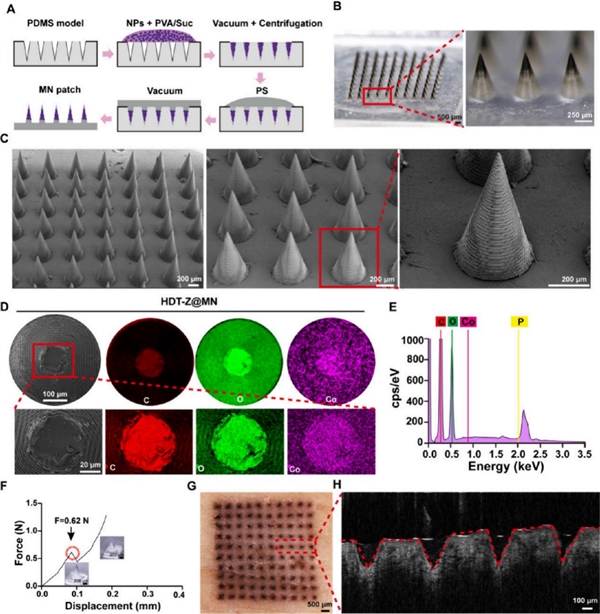
ACS Nano: Microneedle Scalpels Break the Deadlock of Breast Cancer Drug Resistance

The research team led by Professor Li Wei from the School of Pharmaceutical Sciences, Wuhan University, and Professor Song Zhiyin from the College of Life Sciences, Wuhan University, proposed an innovative strategy—a mitochondria-targeted microneedle (HDT-Z@MNs) system that precisely activates the "death switch" of cancer cells to break the doxorubicin resistance deadlock in breast cancer. The team first synthesized DOX-TPP@ZIF-67 nanoparticles (NPs). Cobalt nitrate and 2-methylimidazole were reacted in water to form purple ZIF-67 nanoparticles. Then, DOX modified with triphenylphosphine (TPP) was dissolved in water and added to ZIF-67; the drug was adsorbed by virtue of its pore structure and surface charge, and the reaction was carried out at a pH of about 6.5–7 for 72 hours to obtain hollow DOX-TPP@ZIF-67 NPs.
 
TEM and SEM images showed that the nanoparticles were about 210 nm in size; N₂ adsorption-desorption tests confirmed a mesoporous structure, indicating successful drug loading; a slight decrease in specific surface area and pore volume after drug loading verified the drug loading effect; BJH analysis showed that the pore sizes were concentrated at about 2 nm and 60 nm. The cobalt ion center of ZIF-67 can catalyze the decomposition of H₂O₂, enabling the nanoparticles to propel in H₂O₂-rich environments; enhanced particle movement trajectories, accelerated diffusion rates and increased mean square displacement (MSD) were observed in different H₂O₂ concentrations, demonstrating its role as a "nano-motor" to enhance drug enrichment in tumor and other H₂O₂-rich microenvironments. Drug release was significantly accelerated in acidic environments (pH 5.5) and high H₂O₂ concentrations, indicating pH and H₂O₂ responsiveness, which is suitable for targeted drug release in the tumor microenvironment.
Figure: Characterization of hollow DOX-TPP@ZIF-67 nanoparticle-loaded microneedles (HDT-Z@MNs)
The microneedle array required for the experiment was fabricated by MicroFab Precision's microArch S240 3D printing equipment (precision: 10 μm) and then prepared by PDMS replica molding. This study not only demonstrates the high controllability and safety of the microneedle system, but also proposes a multi-synergistic therapeutic strategy with mitochondria as a new target, providing a new direction for the clinical treatment of breast cancer and other drug-resistant solid tumors.

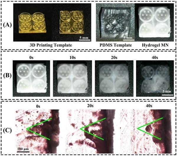
Advanced Science: Drosophila-Bionic Microneedle Patches for Efficient Therapeutic Efficacy

Bacterial infectious stomatitis (BIS) is a common inflammatory disease of the oral mucosa, mainly caused by pathogenic bacteria such as streptococci and staphylococci, clinically characterized by mucosal redness and swelling, severe pain, recurrent ulcers and secondary infections. Although traditional treatments mainly rely on local anti-inflammatory, analgesic and antibacterial drugs, the complex and dynamically changing oral environment often limits drug delivery due to saliva scouring, mucosal barrier and pseudomembrane on ulcer surfaces, leading to unstable therapeutic effects and high recurrence rates. This challenge is driving a shift in oral treatment methods from single drug intervention to the synergistic optimization of structural materials and delivery pathways.
In response to the dynamic characteristics and therapeutic needs of the oral environment, hydrogel materials have attracted extensive attention in local oral treatment in recent years due to their good biocompatibility, high water content and ease of drug loading and modification. By introducing the bionic design concept, hydrogels can be further constructed into microneedle-structured patch devices to achieve stable adhesion and targeted release in lesion areas. A bionic microneedle patch system developed by Professor Fan Zengjie's team from Lanzhou University, inspired by the adhesive structure of Drosophila tarsi, integrates thermosensitive antibacterial hydrogels with bionic sucker design, realizing high adhesion, efficient controlled release and local anti-inflammatory synergistic therapy in the wet oral environment. It has shown significant tissue repair and anti-inflammatory effects in animal models, marking a feasible path for the precise intervention of functional microstructures in the complex oral environment.
Figure: Characterization of microneedle (MN) suckers
The key structure of this novel bionic microneedle patch—the adhesive microneedle array—was fabricated by template processing using MicroFab Precision's Projection micro-stereolithography (PμSL) technology. With a high-precision 3D printing system (microArch S230, precision up to 2 μm), the research team was able to accurately replicate the microstructures of Drosophila claw suckers, achieving geometric reproduction and morphology control of the bionic patch at the substructural scale. This high-resolution and highly repeatable processing capability provides a fundamental guarantee for the bionic functionalization of microstructures, and also opens up new ideas for the large-scale R&D of therapeutic devices with high adhesion and stability in the oral microenvironment.

Chemical Engineering Journal: Mesen Sphere Bandages Accelerate Wound Healing

The therapeutic potential of mesenchymal stem cells (MSCs) in immune regulation and tissue regeneration has been widely verified, especially showing significant advantages in inflammation control and wound repair. Compared with suspended MSCs, mesenspheres with stable structure and stronger paracrine function are becoming an important direction of the new generation of therapeutic strategies. However, the traditional injection-based delivery method has the problems of high invasiveness and low local retention rate, which seriously restrict the efficiency of its clinical application. Therefore, exploring a non-invasive, controllable-release and reusable stem cell delivery platform has become a key task for the technological transformation of regenerative medicine.
Aiming at the technical bottlenecks of current mesensphere dressings in sphere formation efficiency, distribution uniformity, release integrity and repeated administration capability, the latest research from the University of Macau proposed an innovative mesensphere sheet bandage (MSB) solution. Based on polydimethylsiloxane (PDMS) materials with patternable micropores, the dressing not only enables high-throughput self-assembly and uniform release of mesenspheres, but also has the capabilities of repeated application and room temperature storage. Experiments confirmed that the dressing significantly accelerated the wound healing rate (+33%) in mouse models and improved the structural integrity of skin regenerated tissue (+58%) while increasing the secretion level of stem cell factors, showing strong clinical application potential, especially for the treatment of chronic wounds such as diabetic foot ulcers and burns.
For the core manufacturing link of the MSB dressing, the research team used the microArch® S240 equipment (precision 10 μm) to construct a microcolumn array mold, and replicated thousands of high-density microporous structures through PDMS casting. This structure not only realizes the spontaneous aggregation of stem cell suspension into spheres in micro-scale space, but also has the advantages of high consistency, high preparation throughput and simple operation, forming a complete chain from mesensphere preparation and dressing construction to clinical application. MicroFab Precision's high-resolution printing capability and material compatibility have provided key manufacturing support for this innovative achievement, and also laid a solid foundation for the transformation of regenerative medical devices from the laboratory to large-scale clinical application.
Micro-nano 3D printing is not limited to a single structural construction tool. The trends of therapeutic precision, device intellectualization and production customization it brings will profoundly change the traditional R&D paradigm of medical innovation, opening up a new frontier for precision medicine, regenerative medicine and personalized treatment.
 
Disclaimer: This news information is sourced from the internet. If there is any infringement, please contact us for removal. cn@totustec.com

Keywords: